Knee Tendon Diagram - Anatomy Of The Knee - The knee consists of three bones:. Some of the most common symptoms of a torn knee ligament are pain, swelling and, in some cases, an audible snap. The four main ligaments in the knee connect the femur (thighbone) to the tibia (shin bone), and include the following: It is held in place by a ligament at the bottom and a tendon on top. Furthermore, there are several individualized. The knee ligaments connect the thigh and shin bones (femur & tibia) and work together to control how the knee moves to keep it stable and prevent injury.
A diagram of the knee, including ligaments. There are two pairs of ligaments in the knee, collateral ligaments: Bones, cartilage, ligaments, and tendons. The ligament, located in the center of the knee, that controls rotation. Ligaments are elastic bands of tissue that connect bones to each other and provide stability and strength to the joint.
Adolescent Sports Injuries Of The Knee from www.clevelandclinic.org A ligament is a type of fibrous tissue that usually connects two bones. Vectorized and colorized in inkscape, based on image:knee diagram.png. Knee pain could be the result of a problem with any one of these components, or a combination of several. This svg file contains embedded text that can be translated into your language, using any capable svg editor, text editor or the svg translate tool. The knee is the joint where the bones of the lower and upper legs meet. The knee is designed to fulfill a number of functions: One of the most important tendons is the. (the other three are the anterior and posterior cruciate ligaments acl and pcl and the lateral collateral ligament.) the mcl connects the inner (medial) surfaces of the thigh bone (femur) and the shin bone (tibia) and is on the outside of the knee joint.
The knee joint is a complex structure that involves bones.
This svg file contains embedded text that can be translated into your language, using any capable svg editor, text editor or the svg translate tool. The knee joint is a complex structure that involves bones, tendons, ligaments, muscles, and other structures for normal function. It extends from the patella, otherwise known as the kneecap. Some of the most common symptoms of a torn knee ligament are pain, swelling and, in some cases, an audible snap. One of the most important tendons is the. Tendonitis can affect any tendon, but is most commonly seen in the wrist and fingers. Then next one, further down, looks at pain behind the knee. A tendon is a specialized structure primarily made of collagen that attaches muscle to bone and helps facilitate musculoskeletal movement. Damage in even one part can hinder the functioning of the knee. Tendons are elastic tissues made up of collagen. A dislocated kneecap is yet another common knee condition. The patella tendons surround the kneecap and the quadriceps tendons are toward the back of the knee and leg. There are numerous tendons around the knee that also help to stabilize the knee.
Its complexity and its efficiency is the best example of god's creation. The knee is designed to fulfill a number of functions: The anterior cruciate ligament prevents the femur from sliding backward on the tibia (or the tibia sliding forward on the femur). Tendons are elastic tissues made up of collagen. Jumper's knee is inflammation of your patellar tendon, the tendon that connects your kneecap (patella) to your shin bone (tibia).
Thigh Knee And Popliteal Fossa Knowledge Amboss from media-us.amboss.com Portofrei ab 50€, lieferung in 48h! Ligaments join the knee bones and provide stability to the knee: Ligaments are elastic bands of tissue that connect bones to each other and provide stability and strength to the joint. Damage in even one part can hinder the functioning of the knee. A ligament is a type of fibrous tissue that usually connects two bones. When the tendon gives way, you can't move your knee. Jumper's knee is diagnosed by taking a medical history and doing a physical exam. Each of the 6 sections ( bones, connective tissue 1, connective tissue 2, deep muscles, muscles & skin) can be opened up, rotated left or right and viewed more closely.
The largest joint in the body, the knee moves like a hinge, allowing you to sit, squat, walk or jump.
This tendon connects the patella (kneecap) to the tibia. The knee joint is a complex structure that involves bones, tendons, ligaments, muscles, and other structures for normal function. The four main ligaments in the knee connect the femur (thighbone) to the tibia (shin bone), and include the following: A diagram of the knee, including ligaments. The anatomy of the knee consists of bones, muscles, nerves, cartilages, tendons and ligaments. The anterior cruciate ligament prevents the femur from sliding backward on the tibia (or the tibia sliding forward on the femur). Vectorized and colorized in inkscape, based on image:knee diagram.png. When the tendon gives way, you can't move your knee. Three bones meet to form your knee joint: The severity of these symptoms depends on which ligament has been torn. The knee is the joint where the bones of the lower and upper legs meet. One of the most important tendons is the. Cross section of foot nerves 13 photos of the cross section of foot nerves cross section of nerve fiber, foot anatomy nerves, foot nerve pain, human foot nerves, nerve cross section histology, peripheral nerve cross section, spinal nerve cross section, foot, cross section of nerve fiber, foot anatomy.
They are associated with muscles discussed in the section above (see above). There are two pairs of ligaments in the knee, collateral ligaments: The knee is the largest joint in the body who function is to bend (flex) and straighten (extend) in order to allow movement of the body e.g. The largest joint in the body, the knee moves like a hinge, allowing you to sit, squat, walk or jump. Right knee, seen from an angle between anteriorly and laterally.
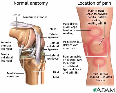
Collateral Ligament Cl Injury Aftercare Medlineplus Medical Encyclopedia from medlineplus.gov Pain above the knee cap (yellow). Around the knee there are two types of tendons. A dislocated kneecap is yet another common knee condition. The anatomy of the knee consists of bones, muscles, nerves, cartilages, tendons and ligaments. There are numerous tendons around the knee that also help to stabilize the knee. The four main ligaments in the knee connect the femur (thighbone) to the tibia (shin bone), and include the following: A ligament is a type of fibrous tissue that usually connects two bones. Knee joint is one of the most important hinge joints of our body.
Acl & pcl found in the middle of the joint.
The ligament, located in the center of the knee, that controls rotation. The kneecap slides along a groove in the femur as the knee bends. Tendons are elastic tissues made up of collagen. Understanding the normal function of the knee joint can help you address some of these common. This first knee pain diagnosis chart focuses on pain at the front of the knee. The four main ligaments in the knee connect the femur (thighbone) to the tibia (shin bone), and include the following: The knee ligaments connect the thigh and shin bones (femur & tibia) and work together to control how the knee moves to keep it stable and prevent injury. There are two pairs of ligaments in the knee, collateral ligaments: Our interactive 3d knee diagram is an informative 360 degree rotating model. It extends from the patella, otherwise known as the kneecap. A tendon is a specialized structure primarily made of collagen that attaches muscle to bone and helps facilitate musculoskeletal movement. Ligaments join the knee bones and provide stability to the knee: It is made up of four main things:


0 Komentar